
Microstructural examination of neutron, proton and self-ion irradiation damage in a model Fe9Cr alloy
Dr Jack Haley, Professor Sergio Lozano-Perez and Professor Steve Roberts, with colleagues from Dalton Cumbrian Facility, CCFE, UK Atomic Energy Authority and UCSB in Santa Barbara, have published a paper in Journal of Nuclear Materials detailing their experiment utilising Transmission Electron Microscopy (TEM).
The TEM was used to compare the microstructural defects produced in a Fe9Cr model alloy during exposure to neutrons, protons or self-ions. It was noted that:
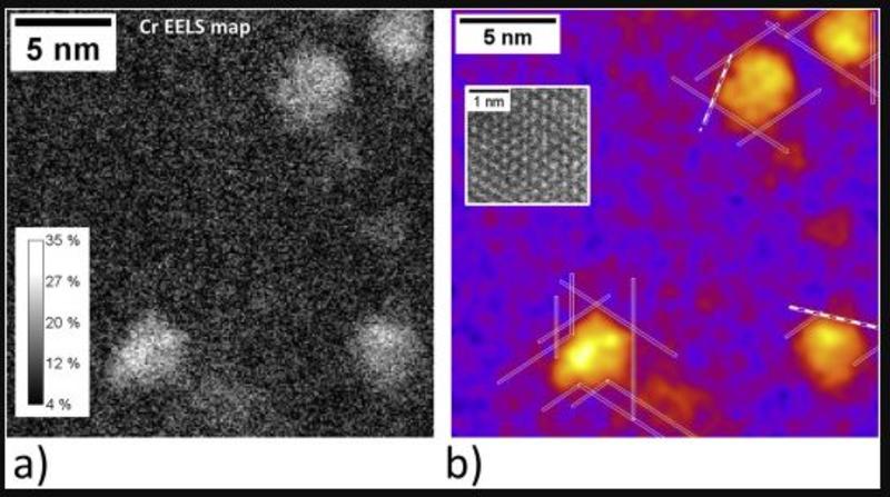
- the proton irradiation redistributes alloyed elements without necessarily requiring a temperature shift to compensate dose-rate;
- vacancy dislocation loops were found to comprise the majority of damage after 2 MeV Fe+ irradition;
- protoon irradiation led to larger cavities, indicating implanted hydrogen might play a role in their formation; and that
- a'-phases were found at density ~9x higher when the dose-rate from neutron irradiation was resolved by a factor of ~3.