
Low-Dose Aberration-Free Imaging of Li-Rich Cathode Materials
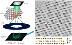
Research by the Peter Bruce Group and collaborators at the Department of Materials as reported in Nano Letters provides an insight to the atomic structure of cathode materials. Imaging the complete atomic structure of materials, including light elements, with minimal beam-induced damage of the sample is a long-standing challenge in electron microscopy. Annular bright-field scanning transmission electron microscopy is often used to image elements with low atomic numbers, but due to its low efficiency and high sensitivity to precise imaging parameters it comes at the price of potentially significant beam damage. In this work they showed that electron ptychography is a powerful technique to retrieve reconstructed phase images that provide the full structure of beam-sensitive materials containing light and heavy elements. Due to its much higher efficiency, beam currents used were reduced down to the subpicoampere range. Electron ptychography also allows residual lens aberrations to be corrected at the postprocessing stage, which avoids the need for fine-tuning of the probe that would result in further beam damage and provides aberration-free reconstructed phase images. Electron ptychography obtains structural information from aberration-free reconstructed phase images in the technologically relevant lithium-rich transition metal oxides at different states of charge. The technique allows to determine the position of the lithium and oxygen atomic columns while amorphization of the surface, formation of beam-induced surface reconstruction layers, or migration of transition metals to the alkali layers are drastically reduced.