
Atomic-scale imaging of polyvinyl alcohol crystallinity using electron ptychography
Polyvinyl alcohol (PVA) is considered to have great potential in medical, pharmaceutical, and packaging applications because of its outstanding biocompatibility, water solubility, low density and relatively low cost.
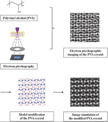
PVA crystallinity, central to the materials properties, has been studied by X-ray diffraction, but two possible crystal structures are mooted. Electron microscopic techniques can potentially image PVA at high resolution - still, it is challenging for conventional electron microscopies because of the relatively low crystallinity of PVA, its severe beam sensitivity, and the poor contrast of light elements.
Electron ptychography makes use of a 4D STEM dataset comprising the intensity in the STEM detector plane recorded as a function of each probe position and has lower sample damage and better phase-contrast compared to traditional techniques. In 'Atomic-scale imaging of polyvinyl alcohol crystallinity using electron ptychography' published in Polymer, the team of researchers, all of whom are based in this department, used electron ptychography to image PVA crystallinity. The reconstructed images, which show good agreement in the unit cell dimension with X-ray diffraction data, can show how the atoms order in the materials, however, deviations from previous models derived from X-ray diffraction are observed.
To interpret the data, the team propose a series of of changes based on previous models to formulate a description of PVA crystal structure. Simulated results from this new model accord well with the experimental images. This study manages to image both carbon and oxygen atoms in PVA, which has not previously been achieved by any conventional method. The results are expected to bring a new and deeper understanding of PVA crystal structure, and illustrate the opportunity presented by this approach for directly imaging molecular order in polymer crystals.