

Near-field ptychography for wide field high-resolution biological imaging
A major challenge in biological imaging is the need to capture large fields of view while preserving structural detail across multiple length scales. Ptychography enables high-resolution imaging over extended areas, though achieving this in practice remains challenging.
In conventional (far-field) ptychography, the detector is positioned sufficiently far from the sample such that the recorded diffraction patterns approximate the Fourier transform of the exit wave. In this regime, the field of view is determined by the probe scan step size and the number of probe positions, and a successful reconstruction of the exit wave typically requires significant overlap (>80%) between the diffraction patterns acquired by adjacent probes. As a result, increasing the field of view is essentially limited by geometric scaling constraints: larger areas require either more probe positions or larger probes, both of which are constrained by experimental and algorithmic limits.
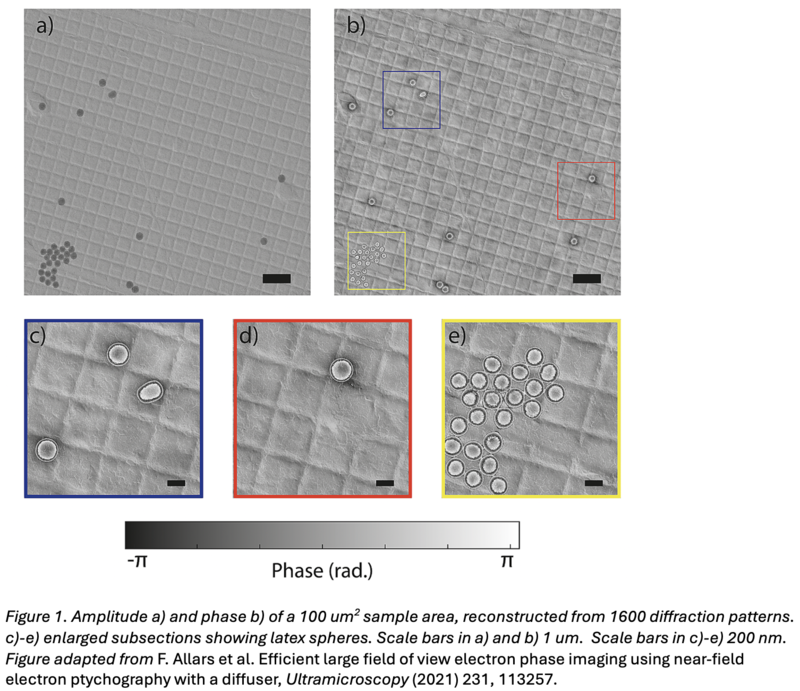
Near-field ptychography offers a fundamentally different approach. Here, the detector is placed closer to the sample, in the Fresnel diffraction regime, where diffraction patterns arise from free-space propagation over a finite distance. In this regime, the recorded intensities exhibit structured interference fringes that encode both amplitude and phase changes over the illuminated region. When combined with structured illumination, this results in significantly richer measurements, in which a single diffraction pattern carries information about a large area of the specimen.
This new imaging approach relaxes the geometrical constraints linking probe size, step size, and detector sampling, enabling the reconstruction of significantly larger fields of view with fewer probe positions, faster acquisition times, and reduced electron dose per unit area. These advantages are particularly important for biological imaging, where radiation sensitivity is a major limitation. Despite this potential, there is currently very little established methodology for applying near-field ptychography to biological specimens.
This project will investigate the fundamental physics of near-field imaging while simultaneously developing new experimental and computational methods. This includes testing structured apertures to maximise phase-amplitude diversity, implementing reconstruction schemes that incorporate near-field propagation, and investigating new data acquisition strategies optimised for wide-field biological imaging.